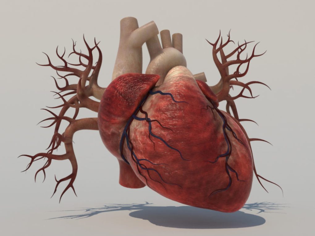
The human heart is only the size of a fist, but it is the strongest muscle in the human body. The heart starts to beat in the uterus long before birth, usually by 21 to 28 days after conception. The average heart beats about 100 000 times daily or about two and a half billion times over a 70 year lifetime.
With every heartbeat, the human heart pumps blood around the body. It beats approximately 70 times a minute, although this rate can double during exercise or at times of extreme emotion. Blood is pumped out from the left chambers of the heart.
It is transported through arteries of ever-decreasing size, finally reaching the capillaries in all the tissues, such as the skin and other body organs. Having delivered its oxygen and nutrients and having collected waste products, blood is brought back to the right chambers of the heart through a system of ever-enlarging veins.
During the circulation through the liver, waste products are removed. This remarkable system is vulnerable to breakdown and assault from a variety of factors, many of which can be prevented and treated.
Heart Anatomy
The heart is a powerful muscle slightly larger than a clenched fist. It is composed of four chambers, two upper (the atria) and two lower (the ventricles). It works as a pump to send oxygen-rich blood through all the parts of the body. A human heart beats an average of 100,000 times per day. During that time, it pumps more than 4,300 gallons of blood throughout the entire body.
Right Ventricle
The lower right chamber of the heart. During the normal cardiac cycle, the right ventricle receives deoxygenated blood as the right atrium contracts. During this process the pulmonary valve is closed, allowing the right ventricle to fill. Once both ventricles are full, they contract.
As the right ventricle contracts, the tricuspid valve closes and the pulmonary valve opens. The closure of the tricuspid valve prevents blood from returning to the right atrium, and the opening of the pulmonary valve allows the blood to flow into the pulmonary artery toward the lungs for oxygenation of the blood.
The right and left ventricles contract simultaneously however, because the right ventricle is thinner than the left, it produces a lower pressure than the left when contracting. This lower pressure is sufficient to pump the deoxygenated blood the short distance to the lungs.
Left Ventricle
The lower left chamber of the human heart. During the normal cardiac cycle, the left ventricle receives oxygenated blood through the mitral valve from the left atrium as it contracts. At the same time, the aortic valve leading to the aorta is closed, allowing the ventricle to fill with blood.
Once both ventricles are full, they contract. As the left ventricle contracts, the mitral valve closes and the aortic valve opens. The closure of the mitral valve prevents blood from returning to the left atrium, and the opening of the aortic valve allows the blood to flow into the aorta and from there throughout the body.
The left and right ventricles contract simultaneously; however, because the left ventricle is thicker than the right, it produces a higher pressure than the right when contracting. This higher pressure is necessary to pump the oxygenated blood throughout the body.
Right Atrium
The upper right chamber of the heart. During the normal cardiac cycle, the right atrium receives deoxygenated blood from the body (blood from the head and upper body arrives through the superior vena cava, while blood from the legs and lower torso arrives through the inferior vena cava).
Once both atria are full, they contract, and the deoxygenated blood from the right atrium flows into the right ventricle through the open tricuspid valve.
Left Atrium
The upper left chamber of the heart. During the normal cardiac cycle, the left atrium receives oxygenated blood from the lungs through the pulmonary veins. Once both atria are full, they contract, and the oxygenated blood from the left atrium flows into the left ventricle through the open mitral valve.
Superior Vena Cava
One of the two main veins bringing deoxygenated blood from the body to the heart. Veins from the head and upper body feed into the superior vena cava, which empties into the right atrium of the heart.
Inferior Vena Cava
One of the two main veins bringing deoxygenated blood from the body to the heart. Veins from the legs and lower torso feed into the inferior vena cava, which empties into the right atrium of the heart.
Aorta
The central conduit from the human heart to the body, the aorta carries oxygenated blood from the left ventricle to the various parts of the body as the left ventricle contracts. Because of the large pressure produced by the left ventricle, the aorta is the largest single blood vessel in the body and is approximately the diameter of the thumb.
The aorta proceeds from the left ventricle of the heart through the chest and through the abdomen and ends by dividing into the two common iliac arteries, which continue to the legs.
Atrial Septum
The wall between the two upper chambers (the right and left atrium) of the heart. Pulmonary trunk: A vessel that conveys deoxygenated blood from the right ventricle of the heart to the right and left pulmonary arteries, which proceed to the lungs.
When the right ventricle contacts, the blood inside it is put under pressure and the tricuspid valve between the right atrium and right ventricle closes. The only exit for blood from the right ventricle is then through the pulmonary trunk. The arterial structure stemming from the pulmonary trunk is the only place in the body where arteries transport deoxygenated blood.


